Table of Contents >> Show >> Hide
- What Is a Bursa (and Why Does Your Knee Have So Many)?
- Knee Bursa Anatomy: The Big Players (Locations You Can Actually Picture)
- What Knee Bursa “Pictures” Usually Show (Without Breaking Copyright or Your Brain)
- Function: What Bursae Do During Real Knee Movements
- When a Bursa Gets Mad: Bursitis Basics (Symptoms, Causes, and Red Flags)
- How Clinicians Tell “Which Bursa” Is the Problem
- Practical Prevention: Keeping Your Knee Bursae Calm
- Experiences: What Knee Bursa Problems Can Feel Like in Real Life (Extended)
- Conclusion
Your knee is basically a high-traffic intersection where bones, tendons, ligaments, and skin all want to pass through
without starting a turf war. The unsung peacekeepers? Bursaetiny, fluid-filled sacs that act like
nature’s “anti-chafe” system. When they’re happy, you never notice them. When they’re angry, you suddenly learn
exactly where the front of your kneecap lives.
This guide walks through knee bursa anatomy, what you’d typically see in “pictures” (diagrams, MRI, ultrasound),
and what each bursa does for your movementplus a longer, real-life-style “experience” section at the end to make it all feel
less like a textbook and more like… an actual knee you have to live with.
What Is a Bursa (and Why Does Your Knee Have So Many)?
A bursa is a small sac lined with synovial-like tissue and filled with a slippery fluid.
Its job is simple: reduce friction and spread pressure between tissues that move against each other.
In a kneewhere bending, squatting, kneeling, running, and pivoting happenfriction management is not optional.
Think of bursae as little “lubrication stations” placed at predictable pinch points: under skin where you kneel,
between tendons and bone where motion is repetitive, and near joint recesses where the knee needs extra glide.
Not everyone has every single bursa as a distinct, separate sac (anatomy varies), but the major ones are common
and clinically important.
Quick knee-bursa takeaway
- Healthy bursae: flat, quiet, basically invisible.
- Irritated bursae: can fill with extra fluid, swell, and hurt (aka bursitis).
- Some bursae are “superficial” (near skin) and some are deep (near tendons/bone or the joint capsule).
Knee Bursa Anatomy: The Big Players (Locations You Can Actually Picture)
There are multiple bursae around the knee, but a handful show up again and again in anatomy charts, imaging reports,
and “why does it hurt right there?” conversations. Below are the most commonly discussed knee bursae and what they do.
1) Suprapatellar Bursa (Suprapatellar Recess)
The suprapatellar bursa sits above the kneecap, deep to the quadriceps tendon and in front of the femur.
In many people, it communicates with the knee joint space and behaves like a joint recess rather than a separate pouch.
Function: It helps the quadriceps tendon and the front of the knee glide smoothly during flexion and extension,
especially as the patella tracks up and down.
Common “picture” clues: On MRI, you may see a fluid-filled recess above the patella; on ultrasound, it can appear
as a compressible fluid pocket that changes with movement or pressure.
2) Prepatellar Bursa (In Front of the Kneecap)
The prepatellar bursa lives between the skin and the front of the patella (kneecap).
It’s superficialmeaning it’s close to the outside worldso it’s vulnerable to repeated kneeling and direct blows.
Function: Lets skin glide over the kneecap when you bend, kneel, or crawl (yes, crawling counts as a knee activity).
Common “picture” clues: When inflamed, swelling is often visibly obvious on the front of the knee,
sometimes described as a “puff” or “bubble” over the kneecap in clinical diagrams.
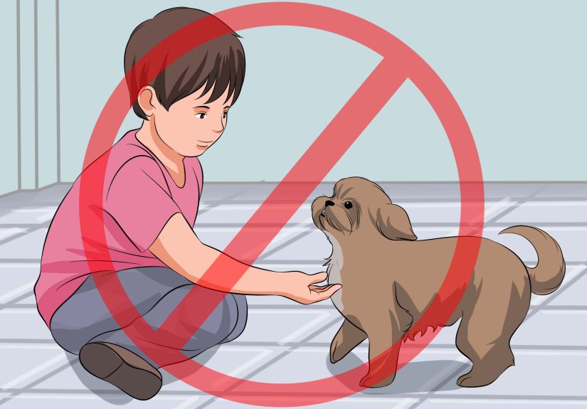
Classic real-world example: Frequent kneeling for work or hobbiesthink flooring, gardening, or sports training.
(This is why prepatellar bursitis is sometimes nicknamed “housemaid’s knee.”)
3) Infrapatellar Bursae (Below the Kneecap)
Under the patella, you’ll hear about two bursae most often:
the superficial infrapatellar bursa (between skin and the patellar tendon) and the
deep infrapatellar bursa (between the patellar tendon and the tibia).
Function: Reduce friction in the “kneeling zone” below the patella and help the patellar tendon glide
during repetitive bending and straightening.
Common “picture” clues: Illustrations often show these as small sacs layered like this:
skin → superficial bursa → patellar tendon → deep bursa → tibia.
On imaging, deep bursitis can be subtle and sometimes overlaps with patellar tendon irritation.
Nickname you might see: The superficial infrapatellar bursa is sometimes associated with “clergyman’s knee”
because kneeling posture can load the area.
4) Pes Anserine Bursa (Inside/Medial Knee, Below the Joint Line)
The pes anserine bursa sits on the inner (medial) side of the knee, typically between the upper tibia
and the tendons of three muscles that insert there: sartorius, gracilis, and semitendinosus.
Function: Helps these tendons glide smoothly over the tibia, especially during walking, stair climbing,
and activities that load the medial knee.
Common “picture” clues: Diagrams often label it a few centimeters below the medial joint line.
On ultrasound, it may appear as a localized fluid pocket; MRI can show fluid and surrounding soft tissue irritation.
Classic real-world example: Medial knee pain in runners, people with knee osteoarthritis,
or anyone with tight hamstrings and repetitive stair usebasically, “life” for a lot of knees.
5) Semimembranosus–Gastrocnemius Bursa (Back of the Knee & Baker’s Cyst Connection)
Behind the knee, a bursa can exist between the semimembranosus tendon and the medial head of the gastrocnemius.
When it fills with fluidoften in connection with joint irritationit may be described as a popliteal (Baker’s) cyst.
Function: Reduces friction in the posterior-medial knee and can act as an overflow “pressure outlet”
when joint fluid increases.
Common “picture” clues: Imaging may show a fluid collection in the popliteal area (behind the knee),
sometimes with a neck-like connection toward the joint space.
What Knee Bursa “Pictures” Usually Show (Without Breaking Copyright or Your Brain)
When people search “knee bursa pictures,” they usually mean one of three categories:
anatomy diagrams, imaging examples (MRI/ultrasound), or clinical comparison images (normal vs inflamed).
Here’s what each tends to highlight so you can understand what you’re looking atno medical degree required.
1) Labeled anatomy diagrams
- Front view diagrams often label the prepatellar and infrapatellar bursae.
- Side (sagittal) view cross-sections show layering: skin, bursa, tendon, bone.
- Medial view diagrams commonly point out the pes anserine bursa below the joint line.
- Posterior view diagrams may show the bursa associated with a Baker’s cyst.
2) Ultrasound images
Ultrasound is great for superficial bursae because it can show a fluid collection close to the skin and can be used dynamically:
press a little, move the knee, and see what changes. It’s also commonly used to guide aspiration or injections when needed.
3) MRI images
MRI is the “zoom out and see everything” option: bursae, tendons, cartilage, menisci, bone marrow, and deeper structures.
If pain is persistent, unclear, or complicated, MRI can show whether a bursa is inflamed and whether something else is also happening.
(If you’re publishing online, use licensed medical illustrations or original diagrams. The key is labeling:
suprapatellar, prepatellar, superficial infrapatellar, deep infrapatellar, pes anserine, and popliteal region.)
Function: What Bursae Do During Real Knee Movements
Knee bursae aren’t “extra parts.” They’re friction managers. Every time you bend your knee, tissues slide.
Without smooth sliding, you get irritation. With enough irritation, you get inflammationand then you get pain.
During kneeling
The prepatellar and superficial infrapatellar bursae take a lot of the pressure and shear.
That’s why people who kneel frequently may develop a visible swelling in front of the kneecap.
During running and stairs
The pes anserine bursa can become irritated by repetitive tendon motion, tight hamstrings,
or altered mechanics (like a knee that collapses inward a bit with each step).
Stairs amplify loadyour knee basically pays extra rent on every step.
During deep flexion (squats, sitting low, certain sports)
The suprapatellar recess helps accommodate patellar tracking and quadriceps glide.
When joint irritation increases fluid, this recess can look more prominent on imaging.
When a Bursa Gets Mad: Bursitis Basics (Symptoms, Causes, and Red Flags)
Bursitis means inflammation of a bursa, often with extra fluid inside it. It can come from mechanical irritation,
direct trauma, systemic inflammation (like gout or rheumatoid arthritis), or infection (especially in superficial bursae).
Common symptoms (varies by bursa)
- Localized swelling (especially prepatellaroften obvious).
- Point tenderness right over the bursa’s location.
- Pain with pressure (kneeling, leaning on the knee) or with certain motions.
- Warmth/redness over the area (more concerning if intense or spreading).
- Reduced comfort with bending rather than true mechanical “locking.”
Common causes
- Repetitive kneeling (prepatellar or superficial infrapatellar).
- Overuse and tendon friction (pes anserine, deep infrapatellar).
- Direct impact (a fall onto the kneecap, sports collisions).
- Underlying joint inflammation (osteoarthritis, inflammatory arthritis).
- Infection (more common in superficial bursae because they’re close to skin and minor breaks).
Red flags that deserve prompt medical evaluation
- Fever or feeling ill along with knee swelling.
- Rapidly increasing redness, heat, or pain.
- Drainage, an open wound, or signs of infection.
- Inability to bear weight, major trauma, or severe limitation of motion.
Friendly reminder: This article is educational. If symptoms are intense, worsening, or you suspect infection,
it’s worth being evaluated by a clinician.
How Clinicians Tell “Which Bursa” Is the Problem
A lot of knee pain is about location + trigger. The knee is a crowded neighborhood, so the fastest path to clarity
is matching your symptom map to anatomy.
Location clues
- Front of kneecap swelling → often prepatellar bursitis.
- Just below kneecap tenderness → infrapatellar bursae or patellar tendon involvement.
- Inner knee pain below joint line → often pes anserine bursitis/tendinopathy.
- Swelling behind the knee → consider a Baker’s cyst (fluid in the popliteal region).
Testing and imaging (typical)
Many cases are diagnosed clinically. Imaging may be used if the diagnosis is unclear, symptoms persist,
or there’s concern for deeper issues. Ultrasound is great for superficial fluid collections; MRI is more comprehensive.
In certain cases, aspiration (drawing fluid) helps evaluate infection or crystal disease.
Practical Prevention: Keeping Your Knee Bursae Calm
You can’t negotiate with biology, but you can stop poking it with a stick. If your day involves kneeling, squatting,
or repetitive bending, bursae appreciate a little common sense.
Habits that help
- Knee pads for work/hobbies that involve kneeling.
- Movement breaks (don’t stay in one kneeling position forever).
- Gradual training for running/stairsload the knee progressively.
- Flexibility + strength (especially quads, glutes, and hamstrings for better knee mechanics).
The goal isn’t to treat your knee like it’s made of glass. It’s to stop treating it like it’s made of indestructible
industrial rubber. Your bursae are small. Your life is not. Compromise is the move.
Experiences: What Knee Bursa Problems Can Feel Like in Real Life (Extended)
Medical diagrams make bursae look neat and politelike perfectly labeled little water balloons tucked into place.
Real life is messier. People don’t show up saying, “Hello, I have inflammation of the superficial infrapatellar bursa.”
They show up saying, “Why does my knee look like it’s smuggling a grape?” or “It hurts every time I go up stairs,
but only on this one weird spot inside my knee.”
The “I didn’t realize kneeling was a sport” moment
A classic prepatellar bursitis story often starts with a totally innocent plan: cleaning floors, gardening, home repairs,
prayer routines, sports drillsanything involving repeated kneeling. The first day, your knee is fine. The second day,
you notice a little tenderness. The third day, the front of the knee looks puffy and feels warm, like it’s trying to grow
its own tiny pillow. The funny part (not funny, but you know) is how quickly you learn the difference between
“knee joint pain” and “superficial, front-of-kneecap pain.” With prepatellar bursitis, the swelling is so localized and
obvious that it can feel like your knee is wearing a tiny inflatable life vest.
The “stairs are suddenly my nemesis” experience
Pes anserine bursitis tends to be sneakier. People often describe it as an ache or sharp tenderness on the inner side
of the knee, a bit below the joint line. It can flare with stairs, getting out of a chair, or long walksespecially if there’s
underlying knee arthritis or a training spike (like adding hills, speed work, or extra step-count goals).
What makes it frustrating is that it doesn’t always show dramatic swelling. Instead, it’s a very specific “thereno,
not the kneecaplower, more inside” pain that can be hard to point out until someone presses the exact spot and you
suddenly become extremely fluent in facial expressions.
The “is it my tendon or my bursa?” confusion
Infrapatellar pain can create a mini detective story because the patellar tendon lives right there too. Some people feel
soreness just below the kneecap with jumping, squatting, or repeated bendingthen wonder if it’s tendon irritation,
bursa irritation, or both. The experience often includes a pattern: it feels stiff when you start moving, improves a bit as
you warm up, then complains later. This is where the “pictures” part of the topic matters in real life: ultrasound and MRI
can help separate “fluid in a bursa” from “thickened tendon” when symptoms overlap.
The “why is there a lump behind my knee?” spiral
A popliteal (Baker’s) cyst experience is its own genre. People notice tightness behind the knee, a fullness that’s more
obvious when standing, or discomfort when trying to fully bend the knee. Sometimes it’s painless and just feels weird.
Sometimes it pairs with underlying joint irritationlike arthritis or a meniscus issueso the cyst feels like the side quest
nobody asked for. The emotional experience can be surprisingly intense because “lump behind knee” is not a fun phrase
to Google at 2 a.m. The good news is that this is often a fluid-related phenomenon rather than something mysterious,
and imaging can usually clarify what’s going on.
The “I can’t tell if this is serious” reality check
Bursitis can be annoying and very treatable, but sometimes it comes with red flagsespecially if a superficial bursa is
infected. People describe the skin feeling hot, looking red, and pain rising quickly rather than gradually. They may feel
unwell overall. That experience is different from overuse irritation; it’s more like the knee is loudly insisting you stop
troubleshooting on your own and get checked out. In those situations, the “bursa” isn’t just a cushion anymoreit’s a
small space where infection can cause trouble.
The “what helped me cope day-to-day” patterns
Across many knee-bursa stories, the most common helpful themes are boring (which is how you know they’re real):
reducing repeated pressure (like using knee pads), taking movement breaks, easing back into activity rather than pushing
through sharp pain, and addressing mechanics (strength, flexibility, and technique). People also tend to do better when
they stop guessing and start mapping their symptoms to anatomy: front-of-kneecap swelling behaves differently than
medial-below-joint-line tenderness. Once you know which “zone” you’re dealing with, the whole situation feels less random.
If there’s a single emotional truth here, it’s this: bursae are tiny, but they can hijack your entire day. The upside is that
because bursae are so location-specific, understanding the anatomy often makes the path forward clearerand gives you
a calmer, smarter way to talk about what you’re feeling with a healthcare professional.