Table of Contents >> Show >> Hide
- Why the 7-Week Ultrasound Matters
- What You Should Usually See at 7 Weeks
- Why You May Not See Everything Yet
- What Happens If the Scan Is Inconclusive?
- When “Let’s Wait” Is Fine, and When It Is Not
- What a “Normal” 7-Week Scan Often Feels Like
- How to Prepare for a 7-Week Ultrasound
- What Not to Assume After One Early Scan
- The Big Picture
- Experiences Related to a 7-Week Ultrasound
- Conclusion
Note: This article is for educational purposes only and is not a substitute for medical care, diagnosis, or treatment.
A 7-week ultrasound is one of those appointments that can feel equal parts magical, nerve-racking, and wildly unfair. You show up hoping for a cinematic debut, and instead the screen may reveal what looks like a tiny sesame seed in a black circle. Welcome to early pregnancy, where a few millimeters can make all the difference and one or two days can completely change what is visible.
If you are around 7 weeks pregnant, a scan often gives helpful information about where the pregnancy is located, whether it appears to be developing on schedule, and whether cardiac activity can be seen. But “often” is not the same thing as “always.” Sometimes everything lines up beautifully on the screen. Other times the sonographer sees less than expected, and that does not automatically mean something is wrong. In early pregnancy, timing is a drama queen.
This guide explains what a 7-week ultrasound usually shows, why a normal pregnancy may still look earlier than expected, when doctors typically repeat the scan, and which symptoms should never be brushed off with a casual “let’s just wait and see.”
Why the 7-Week Ultrasound Matters
In the first trimester, ultrasound is less about adorable baby profiles and more about answering practical medical questions. At this stage, the scan is often used to confirm that the pregnancy is inside the uterus, estimate gestational age, check for a developing embryo, look for cardiac activity, and identify whether there is one embryo or more than one. It can also help explain bleeding, cramping, or uncertainty about dates.
That is why an early pregnancy ultrasound is such a big deal. It is not just a photo opportunity. It is a location check, a timing check, and a reality check wrapped into one slightly awkward appointment.
What You Should Usually See at 7 Weeks
The word usually is doing heavy lifting here, because early ultrasound findings depend on exact dating and the type of scan used. Still, around 7 weeks of pregnancy, these are the common things providers hope to identify.
1. A Gestational Sac
This is often the first structure seen in early pregnancy. It appears as a dark, fluid-filled area inside the uterus. On its own, a gestational sac tells your provider that something is developing in the uterus, but by itself it does not tell the full story. Very early on, a sac may be present before the yolk sac or embryo is visible.
2. A Yolk Sac
The yolk sac is a small circular structure inside the gestational sac. It sounds tiny because it is tiny, but it matters. In early pregnancy, the yolk sac helps nourish the embryo before the placenta takes over. Seeing it is reassuring because it helps confirm that the pregnancy is progressing beyond the earliest stage visible on ultrasound.
3. A Fetal Pole, Also Called the Embryo
The fetal pole is the first visible sign of the embryo. At 7 weeks, it may only measure a few millimeters, which means the ultrasound is basically trying to photograph a poppy seed with career goals. Even so, this little structure gives your provider important information about growth and gestational age. The embryo is usually measured using crown-rump length, a standard early-pregnancy dating method.
4. Cardiac Activity
By about 7 weeks, cardiac activity is often visible, especially on a transvaginal ultrasound. This is one of the most anticipated parts of the visit, and understandably so. People often call it a heartbeat, though in very early pregnancy clinicians may describe it more precisely as cardiac activity. If it is seen, that is encouraging. If it is not seen, context matters enormously, including the embryo’s size and whether the dates are exact.
5. Better Pregnancy Dating
One of the most useful outcomes of a 7-week ultrasound is more accurate dating. If your last menstrual period suggested one date but you ovulated later than average, the scan may shift your due date. That is not the ultrasound being rude. It is the ultrasound being honest.
6. Whether There Is More Than One Embryo
An early pregnancy ultrasound may also reveal twins or another multiple gestation. If your family has a history of surprise twins, this is where the plot twist sometimes arrives.
Why You May Not See Everything Yet
This is the part many people need most. A 7-week ultrasound can be informative, but it is still early. Not seeing everything you expected does not automatically mean miscarriage. Several perfectly understandable reasons can explain a scan that looks “behind.”
Your Dates May Be Off
The most common reason is simple: you may not be as far along as you thought. Pregnancy dating is often based on the first day of your last menstrual period, but ovulation does not always happen on textbook schedule. If you ovulated later than expected, implanted later, or have longer or irregular cycles, a pregnancy that seems 7 weeks by calendar might actually be closer to 5 or 6 weeks in development.
That difference may sound small, but in early pregnancy it is huge. It can be the difference between seeing only a gestational sac, seeing a yolk sac, or seeing an embryo with visible cardiac activity.
The Type of Ultrasound Matters
A transvaginal ultrasound usually gives a clearer picture in early pregnancy than a transabdominal one. That is why many providers prefer transvaginal scanning at 6 to 8 weeks. If the scan is done over the abdomen, especially very early, the image may simply not be detailed enough to show tiny structures clearly.
The Angle of the Uterus Can Change the View
Sometimes the issue is not the pregnancy itself but the viewing conditions. The position and angle of the uterus can affect what is visible. Early pregnancy imaging is not like flipping on a spotlight. It is more like trying to read tiny print through a window from the right angle.
It May Truly Be Too Early
Even in healthy pregnancies, a fetal pole may not be visible until later than expected in some cases. Likewise, an embryo that is visible may still be too small for cardiac activity to be confirmed on that exact day. This is why reputable guidelines use strict measurements and repeat-scan intervals before diagnosing pregnancy loss. Good medicine avoids rushing to conclusions.
There May Be a Real Problem
Yes, sometimes an early ultrasound that shows less than expected can point to miscarriage, a blighted ovum, or an ectopic pregnancy. But that determination usually requires follow-up imaging, serial hCG testing, or both. In other words, one scan is often only one chapter, not the ending.
What Happens If the Scan Is Inconclusive?
If your first scan does not show what your provider hoped to see, the next step is usually not panic. It is follow-up.
In many cases, the plan depends on what was visible:
- If only a gestational sac is seen, a repeat ultrasound is often scheduled about two weeks later.
- If a gestational sac and yolk sac are seen but no fetal pole, a repeat scan is often done after at least 11 days, and many clinicians prefer about two weeks.
- If a small fetal pole is seen but no cardiac activity, a repeat scan is often done in about one week.
This waiting period can feel emotionally ridiculous. You wait for the test, then wait for the answer, then wait to see whether you are allowed to stop waiting. But that repeat scan window exists for a good reason: it helps prevent a viable early pregnancy from being mislabeled too soon.
When “Let’s Wait” Is Fine, and When It Is Not
There is a big difference between a pregnancy that is too early to interpret and one that may be urgent. Call your provider promptly or seek urgent care if you have:
- Heavy bleeding, especially if you are soaking through pads quickly
- Severe or worsening abdominal or pelvic pain
- Lightheadedness, fainting, or feeling like you might pass out
- Bleeding that continues or is paired with significant cramping
These symptoms can happen with miscarriage, but they can also happen with ectopic pregnancy, which can be life-threatening. Early pregnancy deserves gentleness, but it also deserves respect.
What a “Normal” 7-Week Scan Often Feels Like
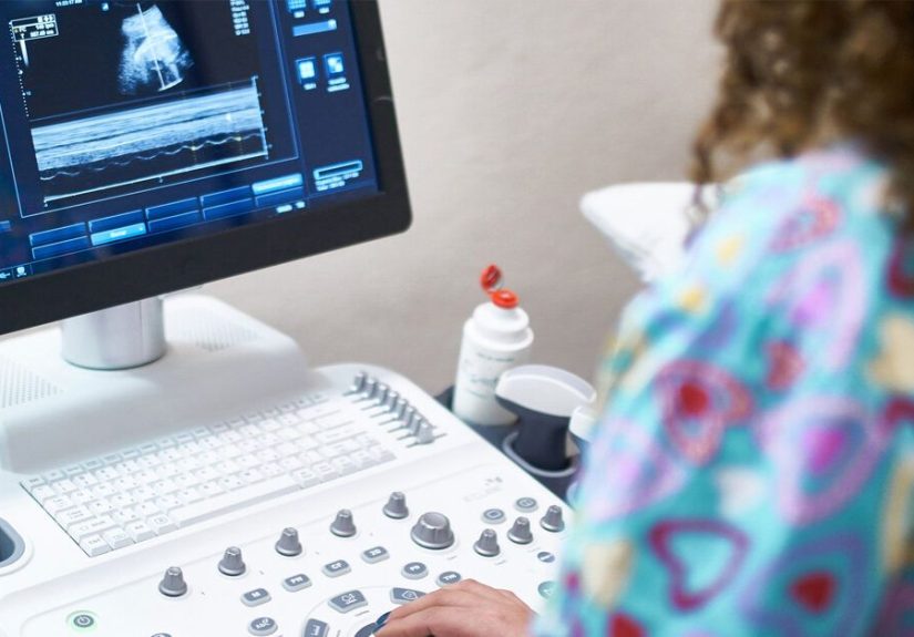
A lot of people imagine the appointment as a dramatic movie moment with instant clarity. In reality, it is usually quieter than that. You may have a transvaginal ultrasound, which can be mildly uncomfortable but is generally quick and well tolerated. The sonographer will capture images, take measurements, and often stay pretty neutral while gathering data. Neutral does not mean bad news. It usually just means they are doing their job and letting the interpreting clinician speak to the results.
That silence, unfortunately, can feel louder than a marching band. If you are nervous, that is normal. If you are trying to decode every eyebrow movement in the room, that is also normal. Pregnancy turns otherwise rational adults into amateur facial-expression detectives.
How to Prepare for a 7-Week Ultrasound
Emotionally
Go in hopeful, but leave room for uncertainty. A reassuring scan can happen. So can an “it may just be early” conversation. Neither outcome says anything about your worth, your choices, or whether you are “doing pregnancy right.”
Practically
Ask whether your scan will likely be transvaginal or abdominal. Bring your date of last menstrual period if you know it. Tell the care team about bleeding, pain, prior ectopic pregnancy, fertility treatment, or irregular cycles. Those details matter because they change how the images are interpreted.
Mentally
Remember that a difference of days matters in early pregnancy. At 20 weeks, a few days barely cause a ripple. At 7 weeks, they can decide whether the embryo looks tiny, whether the scan can confirm cardiac activity, and whether you leave reassured or scheduled for another visit.
What Not to Assume After One Early Scan
Try not to assume these things after a single early ultrasound:
- “No heartbeat today means the pregnancy is over.” Not always.
- “Only seeing a sac means something is definitely wrong.” Not always.
- “My dates cannot be off because I had a period app.” Respectfully, period apps are enthusiastic, not omniscient.
- “If I have spotting, the worst has already happened.” Not necessarily. Spotting in early pregnancy is common, though it should still be reported.
At the same time, do not swing too far in the other direction and ignore symptoms that need evaluation. Calm and cautious can coexist. In fact, they make a great team.
The Big Picture
A 7-week ultrasound is often the first moment pregnancy feels medically real. You may see a gestational sac, yolk sac, embryo, and cardiac activity. You may get a more accurate due date. You may also hear the maddening phrase, “It could just be too early.”
As frustrating as that is, early pregnancy does not always reveal itself on command. Biology has its own schedule, and ultrasound only shows what is physically large enough to be seen on that day, with that equipment, from that angle, at that exact moment. A scan that raises questions is not the same thing as a scan that gives a final answer.
The best next step is usually simple: stay in close contact with your provider, follow the recommended timeline for repeat testing, and get urgent care right away if you develop heavy bleeding, severe pain, or fainting. Early pregnancy is full of suspense, but good follow-up turns suspense into information.
Experiences Related to a 7-Week Ultrasound
The emotional side of a 7-week ultrasound deserves its own section because this appointment is not just medical. It is personal. For some people, the day feels like a celebration. For others, it feels like an exam they did not study for. And for many, it somehow feels like both at once.
One common experience is going in convinced you are exactly 7 weeks, only to hear that the scan looks closer to 6 weeks. That can feel like the room suddenly lost oxygen. Then, a week or two later, the repeat ultrasound shows a growing embryo with clear cardiac activity, and the whole plotline changes. In those cases, the issue was not a failing pregnancy. It was late ovulation, uncertain implantation timing, or a date based on a cycle that did not behave like a textbook cycle.
Another very common experience is spotting before the appointment. Even light spotting can send people into a full internet spiral at 2:13 a.m., which is not exactly the hour of wise decision-making. Some people arrive expecting the worst and end up seeing a reassuring early scan. Others still need follow-up because spotting, while common, does not explain itself. The key emotional truth is that symptoms and scan results do not always match the story you are telling yourself in the waiting room.
People who conceived through fertility treatment often describe the 7-week ultrasound differently. Because their dates may be more exact, the scan can feel less like a mystery and more like a checkpoint. At the same time, that precision can create its own pressure. If the embryo measures behind or the findings are unclear, it may feel even more alarming because there is less room to blame “maybe my dates are off.”
There is also the surprisingly universal experience of expecting the ultrasound image to make instant visual sense. It usually does not. To a trained clinician, the gestational sac, yolk sac, and embryo tell a meaningful story. To everyone else, it can look like abstract modern art with medical billing attached. Many people do not actually feel reassured until the provider explains, in plain English, what is being seen and what it means.
And then there is the waiting. If your scan is inconclusive, the days until the next appointment can feel longer than the entire first trimester. People often say that this is the hardest part: not pain, not the exam, but the not-knowing. If that is your experience, you are not overreacting. Uncertainty is exhausting. It helps to lean on clear medical guidance, limit doom-scrolling, and remember that repeat scans are common because early pregnancy changes fast. Sometimes the answer really is a matter of days.
Conclusion
A 7-week ultrasound can be wonderfully reassuring, confusingly inconclusive, or occasionally an early warning sign that more evaluation is needed. What you should see depends not just on the calendar, but on the exact timing of ovulation, implantation, scan type, and embryo growth. That is why one normal pregnancy may show a yolk sac and embryo with cardiac activity, while another perfectly healthy pregnancy may still look a little too early.
The smartest takeaway is not to expect perfection from a single scan. Expect information. Expect follow-up when needed. And expect that in early pregnancy, a few days can change everything on the screen. That is not a flaw in the process. That is the process.